
Case Report: Customized Partial Scapula Replacement – A Pioneering Approach to Complex Scapula Reconstruction
Reclaiming Shoulder Stability: A New Vision for Scapular Reconstruction When a rare
scapular tumor threatened a 63-year-old patient's shoulder stability and mobility, a meticulously designed customLINK implant offered new hope. This report details the imaging, implant design, and surgical strategy that led to a safe, functional outcome for a complex scapular lesion.

Patient Snapshot: The 63-Year-Old Challenge
A 63-year-old man, 5'10", 126 kg, presented to Parkview Hospital in the United States with a tumor eroding a significant portion of his right scapula. A thorough clinical
evaluation included true AP and lateral radiographs, followed by CT scans to precisely delineate tumor boundaries and assess cortical and cancellous bone stock. These findings supported the decision to pursue a custom-made constrained humeral cup derived from the LINK Embrace standard component.
From Scans to Strategy: Precision Imaging and Planning
CT scans helped define the extent of the lesion and guided the custom implant design. By analyzing cortical and cancellous bone quality, the surgical team was able to anticipate potential difficulties and create a personalized solution aimed at restoring both stability and function.

Tailored Technology: Designing the Custom Partial Scapula
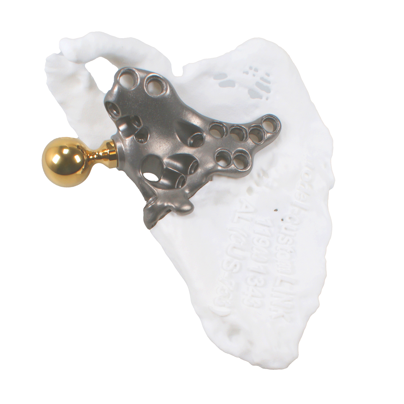
Sophisticated engineering techniques ensured that the implant closely matched the patient's
individual anatomy. TrabecuLink surfaces were incorporated to promote bone contact and
ingrowth. A docking body with integrated flanges allowed the use of either 4.5 mm cortical
screws or 6.0 mm cancellous screws, depending on the intraoperative findings. The partial
scapula component came preassembled with a 25 mm reverse head, streamlining the procedure.
Cutting Edge in the OR: From Preparation to Trial Implant
The surgery began with careful positioning of a custom saw guide aligned with the patient's
anatomical landmarks. This guide was temporarily fixed with 3 mm pins, and resection was
performed along its flat surfaces to remove the tumor-affected bone. A trial implant was then
placed to confirm that the component was fully seated in the bone. Distinct reference marks
could be made at this time to preserve the identified orientation for the final implant.
Securing the Scapula: Final Implant Positioning and Screw Strategy
With the partial scapula prosthesis preassembled, including the head and locking screw, the final implant was aligned according to the position confirmed in the trial. Small pilot holes were prepared using a 3.0 mm K-wire or a 3.2 mm drill. While 4.5 mm cortical screws were the primary choice, 6.0 mm cancellous screws remained an option when bone quality dictated. This was particularly important around the cranial scapula, where four converging screws posed a risk of collision unless carefully selected and aligned. A similar approach was applied to five additional screws, reflecting the adaptable design of the implant.
The Humerus in Focus: LINK EMBRACE System Integration
Once scapular fixation was confirmed, the surgical team implanted a standard humeral stem from the LINK EMBRACE system based on intraoperative assessment. The reverse tray was then secured to the stem, ensuring that its orientation pin was positioned cranially. The fixation screw in the tray was tightened to 3 Nm, verified by an audible click. An inlay was pushed into the tray until another audible click confirmed correct seating. Finally, the humeral cup was coupled to the scapular component using controlled pressure or gentle taps from a designated impactor, and a locking ring was inserted and rotated approximately 180° to achieve secure coupling.

Figure 4 and 5: customLINK implant in situ

A Promising Outlook: Postoperative Evaluations and Early Rehabilitation
Radiographic evaluations demonstrated correct implant geometry and alignment. Early physical
therapy began with an emphasis on securing the reconstruction while promoting residual scapular motion. Although long-term results will require extended follow-up, early clinical results
indicate a promising recovery of shoulder function.
The Future of Scapular Reconstruction: Personalization at its Best
This case illustrates how customized implant strategies can transform care when surgical teams,
biomedical engineers, and imaging specialists work together. By integrating a patient-specific
scapular component with a standardized humeral system, the approach achieved a balance
between innovative technology and practical feasibility. Ongoing monitoring will reveal the
depth of bone integration and functional gains, but early results suggest a brighter future for
patients once considered inoperable.

Read more about our LINK Embrace Shoulder System ...